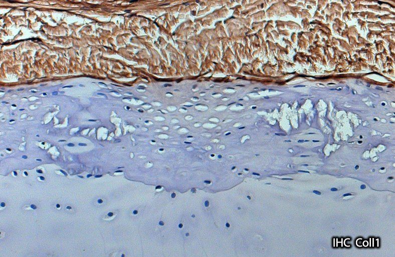
Cartilage or bone? Collagens in tesserae answer an old question
Skeletal mineralization requires an organic framework and in vertebrates collagens form networks guiding the deposition of mineral. In sharks and rays only the skeletal surface calcifies, exhibiting mineralized tesserae sandwiched between a cartilage core and overlying fibrous perichondrium. These two tissues are based on different collagens (Coll II and I, respectively), fueling a long-standing debate as to whether tesserae are more like calcified cartilage (Coll 2-based) or bone (Coll 1-based) in their matrix composition.

In Seidel and Blumer et al.
(Seidel and Blumer et al., 2017. Calcified cartilage or bone? Collagens in the tessellated endoskeletons of cartilaginous fish (sharks and rays). Journal of Structural Biology, 200 (2107): 54-71) we studied the collagenous composition in different tesseral regions, creating a detailed map allowing cross study comparison with our previous study on the development and ultrastructure of tesserae
(Seidel et al. 2016. Ultrastructural and developmental features of the tessellated endoskeleton of elasmobranchs (sharks and rays). Journal of Anatomy 229.5: 681-702) . Moreover, our data indicate a unique potential for tessellated cartilage in skeletal biology research, since it lacks features believed diagnostic for vertebrate cartilage mineralization (e.g. hypertrophic and apoptotic chondrocytes).