Ectopic cartilage mineralizations in elasmobranchs
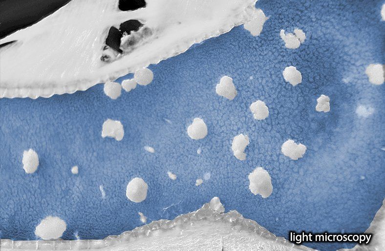
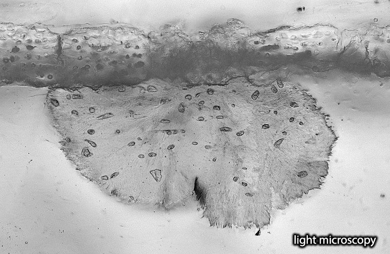
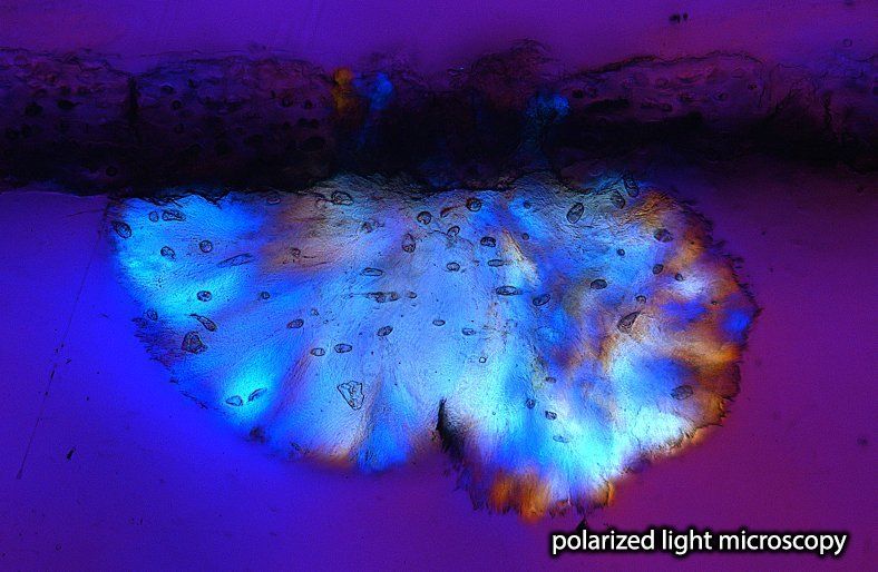
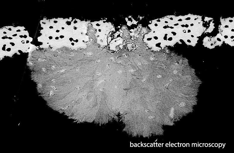
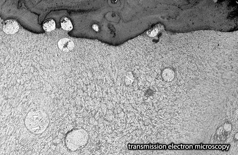
Studying tessellated cartilage in various elasmobranchs species, we noticed in many sections ectopic (located away from normal position) mineralizations. In Seidel et al. 2017 we describe an aberrant type of mineralized elasmobranch skeletal tissue called endophytic masses (EPMs), which grow into the uncalcified cartilage of the skeleton, but exhibit a strikingly different morphology compared to tesserae and other elasmobranch calcified tissues
(Seidel et al., 2017. Ultrastructural, material and crystallographic description of endophytic masses – a possible damage response in shark and ray tessellated calcified cartilage. Journal of Structural Biology 198.1 (2017): 5-18) . EPMs appear to develop between and in intimate association with tesserae, but lack the lines of periodic growth and varying mineral density characteristic of tesserae.
Both tesserae and EPMs appear to develop in a type-2 collagen-based matrix (cartilage), but in contrast to tesserae, all chondrocytes embedded or in contact with EPMs are dead and mineralized. We conclude that a local breakdown of CaP mineralization inhibition processes is a likely cause for EPM formation. The micropetrotic (dead, and filled with mineral) cells within and bordering EPMs suggest that changes in local tissue homeostasis may precede EPM formation. Have a look at our article and see that EPM ultrastructure and crystallography suggest multiple similarities with mineral deposition disorders in elasmobranch but also mammalian cartilage (e.g. chondrocalcinosis associated with osteoarthritis and HDMPs).

