Overcoming the fragility – CT imaging of brachiopod endoskeletons
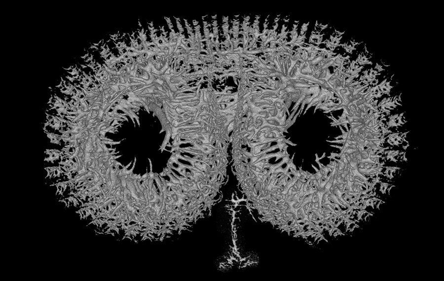
The calcareous shells of brachiopods offer a wealth of informative characters for taxonomic and phylogenetic investigations. In particular scanning electron microscopy (SEM) has been used for decades to visualise internal structures of the shell. However, to produce informative SEM data, brachiopod shells need to be opened after chemical removal of the soft tissue, which occasionally damages the shell. Additionally, skeletal elements of taxonomic/systematic interest such as calcareous spicules, which are loosely embedded in the lophophore and mantle connective tissue become disintegrated during the preparation process. Using micro-computed tomography (micro-CT) allowed us to document the entire fragile endoskeleton of brachiopods for the first time. (Below:
Megerlia truncata 's shells, lophophore and endoskeleton)

Micro-CT is an excellent non-destructive tool for investigating calcified structures in the exo- and endoskeletons of brachiopods. With high quality images and interactive 3D models, our study
(Seidel & Lüter 2014. Overcoming the fragility – X-ray computed micro-tomography elucidates brachiopod endoskeletons. Frontiers in Zoology 09/2014; 11(65):1-15) provides a comprehensive description of the profound differences in shell anatomy, facilitates the detection of new delicate morphological characters of the endoskeleton and gives new insights into the body plan of modern brachiopods.