How do tesserae develop?
It is astounding how little we know about the development of tesserae in elasmobranchs, although early descriptions of tesserae in scientific articles date back to the mid 19th century. Many studies used adult animals to explain the development of tesserae, but also varying species, techniques and changing perspectives made cross-study comparison really hard. As a result, before Seidel et al. 2016, it was not clear "whether tesserae grow in size or number?" or "how tesserae change in shape and mineral density with age?".
(Seidel et al., 2016. Ultrastructural and developmental features of the tessellated endoskeleton of elasmobranchs (sharks and rays). Journal of Anatomy 229.5: 681-702)

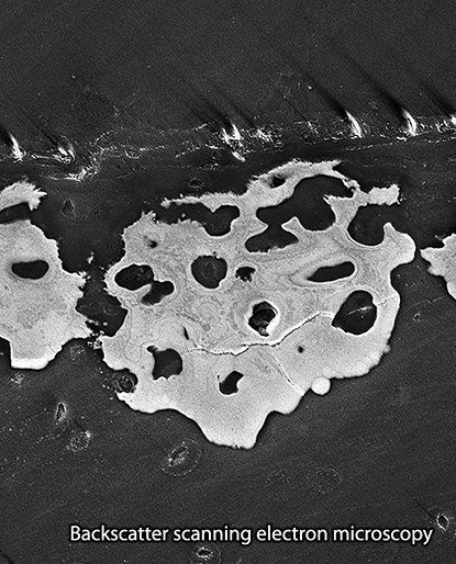
We addressed these questions using biological tissue and material characterization techniques showing that tesserae in size, incorporate living cells and exhibit drastical differences in local mineral density. In particular computed tomography (image above) and backscatter electron microscopy (below) have increased our understanding of the development and ultrastructure of elasmobranch tessellated cartilage, and allowing us to get insights into nature's strategy of tessellating "growing objects".